Normal Biodistribution of Common Radiopharmaceuticals

Nuclear medicine interpretation depends as much on knowing what is normal as on identifying what is abnormal. Every radiopharmaceutical has a predictable physiologic distribution pattern. When you understand this baseline, you instantly recognize when something is off, whether it’s unexpected uptake in an organ, missing activity where it should be, or a shift caused by disease, artifact, or timing error.

Accurate interpretation starts with pattern memory. For example, a bright bowel focus on FDG PET might be physiologic, but if you don’t know FDG’s tendency for variable bowel uptake, you may overcall it as pathology. Likewise, recognizing that Tc-99m sulfur colloid accumulates in the liver, spleen, and bone marrow in fixed ratios prevents false diagnoses of “hepatic cold spots” that are actually physiologic variations.

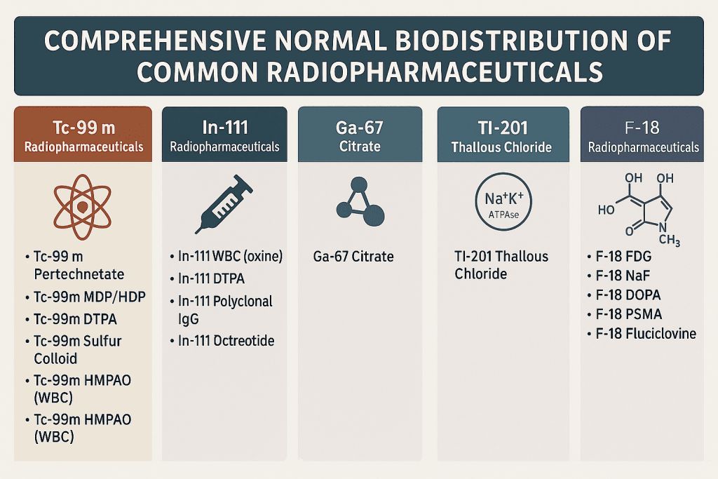
This reference consolidates the normal biodistribution patterns of the most widely used diagnostic radiopharmaceuticals — Tc-99m, In-111, Ga-67, Tl-201, and F-18 — drawn from the Society of Nuclear Medicine and Molecular Imaging (SNMMI) Procedure Standards and leading textbooks.
It’s designed for daily clinical use, teaching, and board exam review.


Tc-99m Radiopharmaceuticals

AgentMechanismNormal Uptake Sites
Tc-99m PertechnetateActive transport (iodide analog)Thyroid, salivary glands, gastric mucosa, choroid plexus, nasopharynx, esophagus, testes, bladder
Tc-99m MDP / HDPChemisorption to hydroxyapatiteAll bones (especially axial skeleton), growth plates, joints, bladder
Tc-99m DTPAGlomerular filtrationKidneys (cortex), ureters, bladder
Tc-99m MAG3Tubular secretionKidneys (parenchyma), ureters, bladder
Tc-99m Sulfur ColloidPhagocytosis by RESLiver (homogeneous), spleen, bone marrow
Tc-99m HMPAO (WBC)Leukocyte labelingLiver, spleen, bone marrow (no bowel activity)
Tc-99m DISIDA / HIDAHepatic excretionLiver, gallbladder, biliary tree, small bowel
Tc-99m RBC (tagged)Intravascular compartmentHeart, blood pool, liver, spleen
Tc-99m SestamibiPassive diffusion, mitochondrial uptakeMyocardium, salivary glands, thyroid, liver, kidneys, bowel, bladder
Tc-99m TetrofosminPassive diffusionMyocardium, liver, kidneys, bladder

In-111 Radiopharmaceuticals

AgentMechanismNormal Uptake Sites
In-111 WBC (oxine)Phagocytosis by neutrophilsLiver, spleen, bone marrow (no bowel or kidney activity)
In-111 DTPACSF distributionCSF spaces (ventricles, cisterns), bladder
In-111 Polyclonal IgGFc receptor bindingLiver, spleen, bone marrow, blood pool
In-111 OctreotideSomatostatin receptor bindingSpleen, liver, kidneys, thyroid, pituitary, bowel, bladder

Ga-67 Citrate

MechanismNormal Uptake Sites
Binds to transferrin, lactoferrin, siderophoresLiver, spleen, bone marrow, salivary glands, nasopharynx, lacrimal glands, breasts, lungs (mild), bowel (variable), kidneys (early), bladder (early)

Tl-201 Thallous Chloride

MechanismNormal Uptake Sites
Na⁺/K⁺ ATPase pump (potassium analog)Myocardium, skeletal muscle, liver, kidneys, testes, salivary glands, thyroid, spleen, bowel (mild), bladder

F-18 Radiopharmaceuticals

AgentMechanismNormal Uptake Sites
F-18 FDGGlucose analog via GLUT transportersBrain (high), myocardium (variable), liver, kidneys, bladder, salivary glands, thymus (children), GI tract (variable), brown fat (neck, supraclavicular), testes, endometrium (cyclical), breasts (lactating), muscles (active), vocal cords (talking), lymphoid tissue
F-18 NaFChemisorption to boneSkeleton, bladder
F-18 DOPAAmino acid transportBrain, pancreas, adrenal medulla
F-18 PSMABinds to PSMA receptorSalivary glands, kidneys, liver, bowel, bladder
F-18 FluciclovineAmino acid transportLiver, pancreas, muscle, bladder

Clinical Tip
  • Recognize asymmetric or unexpected intensity in a physiologic site; it’s often your first clue for pathology.
  • Always correlate biodistribution with patient preparation, injection site, and timing.
  • Reviewing these patterns regularly improves speed and accuracy in board and clinical reporting.

References:
  1. Mettler FA, Guiberteau MJ. Essentials of Nuclear Medicine and Molecular Imaging. 7th ed. Philadelphia: Elsevier; 2019.
  2. Cherry SR, Sorenson JA, Phelps ME. Physics in Nuclear Medicine. 4th ed. Philadelphia: Elsevier Saunders; 2012.
  3. Society of Nuclear Medicine and Molecular Imaging. Procedure Standards and Guidelines. [Internet]. SNMMI; 2025 [cited 2025 Oct 20]. Available from: https://www.snmmi.org/ClinicalPractice
  4. Palestro CJ. Radionuclide imaging of infection: In search of the grail. J Nucl Med. 2009;50(5):671–673.
  5. Love C, Palestro CJ. Radionuclide imaging of infection. J Nucl Med Technol. 2004;32(2):47–57.
  6. Hoffer PB. Gallium: mechanisms. J Nucl Med. 1980;21(7):667–670.
  7. Kostakoglu L, Goldsmith SJ. 18F-FDG PET evaluation of the response to cancer therapy. Semin Nucl Med. 2003;33(4):328–341.
  8. Delbeke D, Coleman RE, Guiberteau MJ, et al. Procedure guideline for tumor imaging with 18F-FDG PET/CT 1.0. J Nucl Med. 2006;47(5):885–895.
  9. Silberstein EB, Ryan J. Prevalence of physiologic and benign variants on whole-body FDG PET imaging. Radiographics. 2001;21(2):545–559.
  10. Bombardieri E, Aktolun C, Baum RP, et al. 111In-pentetreotide scintigraphy: procedure guidelines for tumor imaging. Eur J Nucl Med Mol Imaging. 2003;30(5):BP146–BP155.

Explore more: Visit Masters of Nuclear Medicine for board-style MCQs, mock exams, and system-based volumes that consolidate these principles for daily practice.

Leave a Comment

This site uses Akismet to reduce spam. Learn how your comment data is processed.

Scroll to Top

Discover more from Masters of Nuclear Medicine

Subscribe now to keep reading and get access to the full archive.

Continue reading